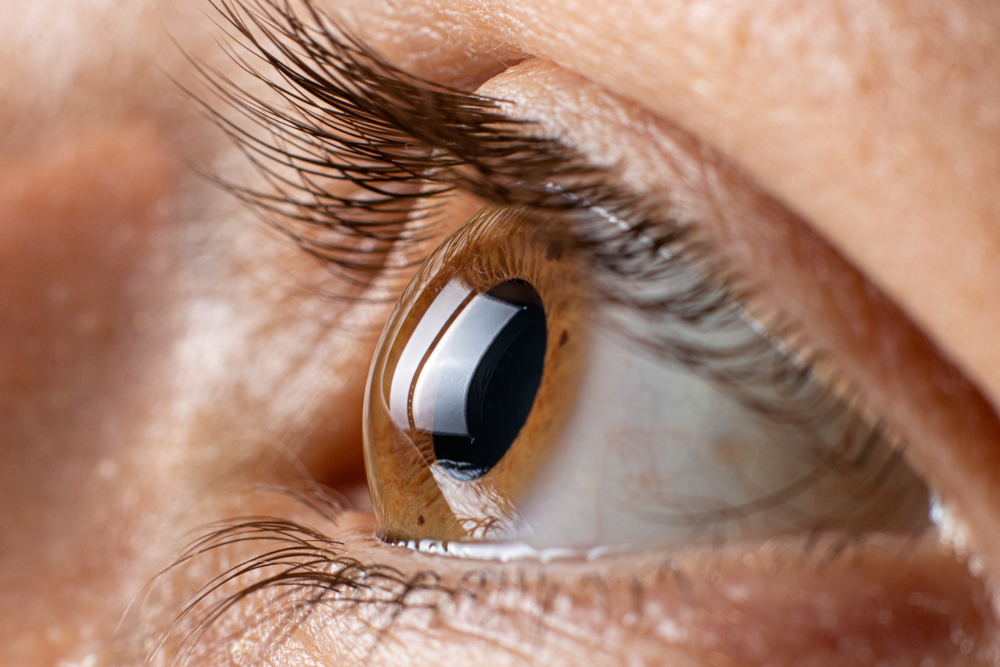
Keratoconus is an eye disorder that affects the cornea, the clear front surface of the eye, causing it to thin and bulge outward into a cone-like shape. This distortion of the cornea leads to blurred vision and sensitivity to light, among other symptoms. It can affect one or both eyes and is most commonly diagnosed in young people or in their late teens.
What are the Causes and Symptoms of Keratoconus?
The exact cause of keratoconus is unknown. However, it's believed to be a combination of genetic, environmental, and hormonal factors. Some studies suggest that keratoconus can run in families, and if a parent has it, there's a one in ten chance their child will have it too. Allergies and frequent eye rubbing also appear to be associated with keratoconus, although it's not clear whether they are causes or effects of the condition.
The symptoms of keratoconus can vary in severity and pace of progression. In the early stages, symptoms can be mild and include slightly blurred or distorted vision, increased sensitivity to light and glare, and frequent changes in eyeglass prescription. As the disease progresses, symptoms may include increased blurring and distortion of vision and increased nearsightedness or astigmatism (irregular curvature of the eye's front surface). In severe cases, the cornea can become scarred, causing further vision impairment.
How is Keratoconus Diagnosed?
Diagnosing keratoconus involves a series of eye examinations, often over several visits to your optometrist. Initially, your eye doctor may notice irregularities in your cornea during a routine eye exam. If keratoconus is suspected, further tests, such as corneal topography (which maps the surface of the cornea) and optical coherence tomography (which provides a detailed view of the cornea's layers), may be conducted.
Another diagnostic tool is the slit-lamp examination, which involves shining a thin sheet of light into the eye while the doctor looks through a microscope. This test allows the doctor to look for signs of keratoconus such as corneal thinning, Fleischer rings (iron-colored rings surrounding the cornea), and Vogt's striae (stress lines caused by corneal thinning).
What are the Treatment Options for Keratoconus?
Treatment for keratoconus depends on the severity of the condition and how quickly it is progressing. In the early stages, vision problems can often be corrected with prescription glasses or soft contact lenses. As the condition progresses and the cornea becomes more irregular in shape, rigid gas permeable (RGP) contact lenses may be recommended. These lenses are more durable and maintain their shape, providing a clearer vision than soft lenses.
Another option is hybrid contact lenses that have a rigid center with a softer ring around the outside for comfort. In more advanced cases of keratoconus, scleral lenses (large-diameter contacts that vault over the cornea and rest on the white part of the eye) may be used.
For those who cannot tolerate contact lenses or for whom lenses and glasses no longer provide acceptable vision, surgical options may be considered. These include corneal inserts, corneal collagen cross-linking, and in severe cases, corneal transplantation.
Embracing Life with Keratoconus
Living with keratoconus can be challenging, but with early diagnosis and the right treatment, most people can lead normal lives. It's important to have regular eye exams, particularly if there's a family history of the condition, and to seek specialist advice if you notice changes in your vision. The outlook for people with keratoconus has improved significantly in recent years, thanks to advancements in contact lens technology and surgical procedures.
For more information on keratoconus, its causes, symptoms, and treatment options, visit Hilltop Eye Center at our office in Liberty, Missouri. We value your experience, take the time to answer questions and ensure you understand all your options for optimal eye health. Please call (816) 781-0500 to schedule an appointment today.


